Trigger Finger and Trigger Finger Release Surgery

What is Trigger Finger and How is it Diagnosed?

Trigger finger, called stenosing tenosynovitis by doctors, is a condition where the finger tends to get locked in place when you are bending it toward the palm. Most of the time, your family doctor will examine you and note the problematic symptoms. This condition is more common among people with rheumatoid arthritis or diabetes. The doctor usually refers you to an orthopedic hand specialist when the finger gets stuck or clicks and is not able to straighten.

The symptoms of trigger finger occur without injury most of the time, or they may follow a period of heavy hand usage. A tender lump can develop in your palm and there may be swelling. The main symptom is inability to straighten the finger without pain and the finger will catch or pop. The stiffness of trigger finger tends to worsen after periods of inactivity and many find that it is worse in the mornings.

Often times, trigger fingers can be treated with a steroid injection in the office. If the finger continues to catch or lock after a steroid injection, the treatment for this condition becomes surgery to open up the flexor tendon sheath in order to eliminate the catching or locking.

What Do I Need to Do Before Trigger Finger Release Surgery?

Before you undergo this procedure, your orthopedic hand specialist will discuss with you exactly what will happen before, during, and after the procedure. You can prepare yourself by asking questions to help you be well informed so you can go ahead with the consent for surgery and sign the necessary forms.

The operation is done under local or regional anesthesia, which means you will be awake and alert during trigger finger release surgery. Your hand will be totally numb, however, allowing your surgeon to operate painlessly.

What is Involved with Surgery?

Once the anesthetic has taken effect, your orthopedic hand specialist will make a 2cm incision into the palm of your hand so he or she can get to the tendon. The surgeon then will release the tendon by making a small incision in the first annular pulley of the tendon sheath. Once this has occurred, you may be asked to move your fingers and to make a fist.

Don’t worry, because of the anesthesia, this won’t hurt. Once the doctor is sure the tendon is properly released, he will close the incision and cover the wound area with a bandage.

What Can I Expect After the Trigger Finger Release Surgery?

It may take several hours before the feeling comes back in your hand so you must be careful not to bump or knock the area. You may need to take pain medication for a few days following this operation as well.

If you have general anesthesia, you will need to rest in a recovery room until the effects of it have worn off. The nurse or doctor will give you follow-up care advice and a follow-up appointment. You will need to keep your dressing and stitches clean and dry for around ten days and then they will be removed. It usually takes about three or four weeks to fully recover from trigger finger release surgery, so it is important that you follow your orthopedic specialist’s advice during this time.

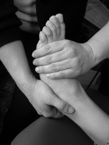
When Can Physical Therapy Help?

You may be wondering if or not you are a candidate for physical therapy (PT), or question whether physical therapy can help your ailment. This is an easy question for most physical therapists to answer: Yes, most people benefit from some form of physical therapy, whether their problem is simple (like an ankle sprain) or more severe (like a serious neurological disorder. At Orthopedic Specialists of Seattle, our physical therapists describe PT as conservative treatment that addresses the healing, management, and prevention of disabilities and injuries.

Physical therapy uses non-invasive and non-medical techniques and tools to help you improve total body function. Our physical therapist focuses on relief of pain, promotion of healing, restoration of function and movement, and adaption and facilitation associated with the injury involved. PT also focuses on body mechanics training, wellness, and fitness so you can improve your quality of life. Our physical therapist uses exercise, cold therapy, heat treatments, electricity, and therapeutic massage to achieve the goals of restoring maximum functioning to each individual patient. … read more

Biceps Tendon Ruptures

Have you felt a pop in your arm and been concerned that you may have torn your biceps?

You are not alone – this common injury affects thousands of Americans every day, with the typical tear occurring in males 30-50 years old, often with a distinct tearing feeling or even an audible “pop.”

These tears often cause significant bruising and loss of function and tend to do poorly without surgical reattachment of the torn tendon. Fortunately, there have been significant advances in the understanding of the tear and proper repair within the last couple of years that not only allows for a significantly stronger repair, but also allows for earlier recovery through minimally invasive treatments.

What is a biceps tear?

It is important to understand a bit of anatomy before delving into the specifics of the biceps tear rupture specifics. The biceps tendon has two attachments at the shoulder and one attachment at the elbow. The biceps tendon is not only important for elbow flexion, but also forearm supinaton –rotation of the forearm that allows us to open up a door or hold our hand out for change. Rupture of the biceps tendon at the shoulder or elbow will cause dysfunction in both functions.

Proximal Biceps Tendon Rupture

The typical injury to the upper end of the biceps is where the biceps tendon ruptures from its attachment at the shoulder joint, specifically at the superior labrum of the glenoid bone. Typical symptoms include shoulder pain, bruising, and often a bulging, shortened biceps muscle known as a “Popeye muscle.” Often patients who tear their biceps tendon describe preexisting pain at the front of the shoulder and pain with shoulder movement called biceps tendonitis. Biceps tendonitis can often be prophylactically treated to prevent or minimize biceps rupture, often through physical therapy, steroid injections or shoulder arthroscopy to debride or repair the tendon.

With Proximal Biceps tendon rupture, many active patients notice a slight loss of strength and a significant cosmetic change in their arm with the bulging muscle and many benefit from repair. I perform the proximal biceps tendon repair as an outpatient procedure and is often quite successful, typically allowing for full return to previous activity.

Distal Biceps Tendon Rupture

Injury to the distal biceps occurs when the biceps tendon is being flexed against a significant force. There is typically a “pop” or a tearing sensation followed by bruising and retraction of the biceps muscle. In a significant percentage of patients, this initial episode is followed by a reasonable return to activity over the next several weeks, delaying care. Active patients often notice pain, weakness and difficulty in twisting activities such as opening a door. Elbow flexion is somewhat preserved due to the presence of the brachialis muscle, which is quite strong and rarely injured.

Treatment for distal biceps tendon rupture includes prompt early diagnosis as the retracted muscle and tendon quickly scars into its retracted position. For the vast majority of patients, surgical reattachment is recommended and can be done as an outpatient procedure.

New Repair Treatment Technique

I perform a newer technique of Biceps Tendon repair, which includes performing the surgery through a very small incision, typically 2 centimeters at the elbow. The smaller incision is utilized due to a newer biceps button technique that provides not only stronger fixation and proper tension, but also earlier recovery due to the strength of the repair and the limited nature of the incision and dissection.

Chronic biceps tendon ruptures (typically over 3 months old) might require larger dissection, due to the retracted scarred nature of the tear. They can occasionally require the use of additional tendon graft, but still utilize newer more-reliable fixation techniques that allow for quicker rehabilitation.

All in all, advances in Biceps tendon tear repair allows for early return to activity and strength. The key to proper treatment includes early identification and diagnosis and is quite rewarding to help patients return to their pre-injury state of function.

Do not hesitate to contact us for further questions or for a prompt evaluation.

Read the original article on by our very own Dr. Scott Ruhlman. He performs the new surgical technique that allows for stronger repair and earlier recovery.

ACL Tears and Knee Arthroscopy

What is the ACL?

The most commonly injured ligament of the knee is the anterior cruciate ligament or ACL. The risk of injury is greater among those who participate in high-risk sports like football, basketball, soccer, and skiing. Around half of ACL injuries occur in combination with damage to the meniscus (cartilage pad), other ligaments, or other structures.

Most of the time, injury to the ACL occurs with pivoting, sidestepping, awkward landings or difficult cutting movements.

The knee is a hinged joint that is held together by four ligaments, one of them being the ACL. This structure runs diagonally in the middle portion of the knee to prevent the lower leg bone (the tibia) from sliding out in front of the thighbone (the femur). The ACL also serves to provide rotational stability of the knee.

What is Knee Arthroscopy?

One type of knee surgery is arthroscopy, a common surgical procedure in which a joint is viewed using a tiny camera. This camera looks into the knee through a small lens and projects an image on a TV monitor to allow the orthopedic specialist a clear view of what is inside the joint space.

An arthroscopic knee procedure allows the surgeon to diagnose and treat the knee injury at the same time. According to the American Orthopedic Society, more than 4 million knee arthroscopies are performed each year. This is a safe and effective way to treat the torn ACL.

Almost all arthroscopic knee surgical procedures are done on an outpatient basis. Knee arthroscopy is most commonly used for reconstruction of the torn ACL, trimming away pieces of torn cartilage, removing loose bone fragments or pieces of cartilage, and removing inflamed synovial tissue.

Arrival
Your surgery center or hospital facility will contact you with specific details about your appointment. You will be asked to arrive an hour or two before the procedure and not to eat or drink after midnight the night before.

Anesthesia
Once you are at the facility where the knee arthroscopic procedure will take place, a member of the anesthesia team will see you. Knee arthroscopy or ACL surgery is usually performed under regional, or general anesthesia. The anesthesia professional will help you decide which method is best for you.

Procedure
The orthopedic specialist will make three small incisions in your knee. A sterile solution will be used to irrigate the knee joint to wash away any cloudy fluid. This helps your surgeon see your knee clearly and in great detail. If surgical treatment is required, your orthopedic surgeon will insert tiny instruments through another incision. These instruments might be trimmers, scissors, and motorized shavers.

ACL tears are repaired by the use of substitute grafts, usually made of tendon. The graft tissues often involve autograft, meaning it comes from the patient. Tendons commonly used are the patellar tendon or the hamstring tendon. During the procedure, the orthopedic specialist will often drill small bone tunnels into the tibia and femur to place the ACL graft in the same position as the torn ACL. The graft is held under tension and is fixed in place using screws, washers, posts, or staples.

Recovery
Rehabilitation after ACL surgical reconstruction with arthroscopy is much faster than recovery from traditional open knee surgery. You will need to follow your orthopedic specialist’s instructions carefully. Be prepared for some swelling, so you will need to keep your knee elevated as much as possible the first couple of days after the procedure. Ice is usually recommended by the surgeon to take down swelling and help with the pain.

Dressing Care
Once the procedure is over, the doctor will cover your knee with a sterile dressing and you are to keep the incisions and this dressing clean and dry. Your orthopedic specialist will advise you on bathing and when to change the dressing.

Bearing Weight
After arthroscopic knee surgery to reconstruct the ACL, you will likely need crutches or some other assistive device for the first few days to prevent excessive swelling and bleeding into the knee. Your surgeon will tell you when it is safe to bear weight on your leg and foot.

Driving
Typically, you must wait 1 to 3 weeks before driving, depending on your doctor’s orders. The orthopedic specialist will base this decision on the knee that is involved, whether your car is an automatic or stick shift, the nature of your procedure, what type of medications you are taking, and how well you can control your knee.

Rotator Cuff Tears


Many Americans suffer rotator cuff tears and they are a common cause of pain and disability. When you tear your rotator cuff, you weaken your entire shoulder making daily activities more difficult. Just raising your hand up to comb your hair could cause serious pain. Read on to find out what makes up the rotator cuff, who is at risk for this type of injury, what are the symptoms of a tear, and how a rotator cuff is treated. … read more

Tell us how we did!
close slider