Barefoot Running

Who doesn’t want to run like a Kenyan? The speed, endurance, and efficiency of these elite distance runners is the stuff of legend, and those in the running community have tried to glean some insight as to what makes these African runners such a powerful force in marathon running. One of the obvious starting points is to analyze the biomechanics of the running stride and see if there are efficiencies inherent to the Kenyan athlete.

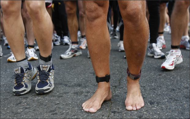
Of course, this has been done with more than one researcher noting one glaring observation: Kenyan runners do not wear shoes. They grow up, play, and often train barefoot. Could this be the secret to running faster? Certainly, some people think that it is. The barefoot running trend has gained a steady following over the past few years. But as the barefoot running contingent has grown, so have its detractors. Let’s take a closer look.

Barefoot Running

Advantages of Barefoot Running

First of all, most barefoot runners do not run in their bare feet. Even the fanatics realize that the roads and trails contain many hazards such as broken glass, nails and rocks that could cause potential injury or discomfort to the feet.

Instead, they use minimalist running shoes, a type of sneaker designed to mimic the barefoot condition in terms of biomechanics. Typically, these shoes are lightweight and feature a thin sole without the large heel cushion found in traditional running shoes.

Biomechanically, the research has shown that barefoot running eliminates or minimizes the heel strike during running. The runner attempts to absorb the impact of body weight by landing with the foot flat or slightly on the ball of the foot. This allows the lower leg and foot to distribute the body weight over a larger surface area. The heel strike found in those wearing traditional running shoes, called shod runners, creates a condition where the full force of impact is driven through the heel, and ultimately the heel cushion of the shoe.

Proponents of barefoot running claim reduced injuries as a result of this change, although there is not much research available to support this claim. One claim that does seem to be supported in the medical literature however, is that of reduced energy consumption while running barefoot.

Simply put, barefoot runners should not fatigue as quickly as shod runners. This would be a great advantage to distance runners and racers who want to attain peak performance or even achieve a personal best during local road races.

The finding is interesting as stride frequency and mechanical work were higher in barefoot runners, indicators which would lead one to believe that the runner would consume more energy. However, the cushioning material in a running shoe absorbs a considerable amount of energy in the shod runner. Energy that would otherwise be used to propel the runner forward is lost in the sneaker. Think of the traditional running shoe like a Cadillac. It gives a smooth ride, but not too efficient.

Disadvantages of Barefoot Running

The obvious risks associated with barefoot running such as puncture wounds can be mitigated with the use of a minimalist running shoe. With this type of footwear, much of the biomechanical adaptations which proponents claim as advantageous are maintained, i.e. reduced heel strike and improved efficiency.

However, there are other reasons why someone may not want to run barefoot. Without a traditional running shoe, the runner lands with a flat foot instead of the traditional heel strike seen in shod runners. This increases the pressure on the bones of the forefoot, which are quite a bit more fragile than the heel.

Over time and with high mileage, a runner could develop a stress fracture, a small break in one of the forefoot bones. This would sideline a runner for several weeks at best, and could become more severe if ignored. Proponents claim that barefoot running is more natural and that we as humans evolved in a way that makes the barefoot method more efficient. But cavemen rarely put in thirty plus miles per week.

The bottom line is that there has not been enough research performed to advocate one method or the other. More studies need to be conducted, and we need to be open-minded about the results. With the barefoot trend steadily gaining a following, the research is sure to follow.

In the meantime, let’s go back to our elite Kenyan marathoners. In an environment where every conceivable advantage is sought and analyzed, these athletes all wear running shoes in competition. Maybe shod running is biomechanically advantageous, or maybe the cumulative effect of pavement on flesh for 26.2 miles eliminates the inherent advantage of running barefoot.

Dr. Mark Reed is an orthopedic surgeon specializing in foot and ankle surgery in the Seattle metro area. He can address all of your questions regarding barefoot running as well as any other foot and ankle conditions.

barefoot running photo

5 Common Hockey Injuries

Hockey photo

The regular NHL season is well underway and the Seattle Thunderbirds are respectively improving their stats with their recent win over the Vancouver Giants at ShoWare Center.

In the regular season as well as the off season, players experience a variety of sports-related injuries as a result. Ice hockey is a contact sport where the players and the puck move at high speeds, so when players run into each other or objects, great force is used. This is why hockey is considered a collision sport. Injuries are fairly common, but efforts can be made to avoid them with training and proper equipment. Listed below are five common injuries that can occur while playing hockey.

AC Joint Injury

The acromioclavicular joint, or AC joint, is one of the joints in the shoulder responsible for motion and stability. The ligaments that hold the AC joint together can be torn through sudden impact to the shoulder, which can cause separation to occur in the AC joint. This sudden impact can happen in hockey when players skating at high speeds collide with one another or into a rigid surface. Swelling, bruising, pain, and motion range loss are all symptoms of AC joint separation. There may also be visible bumps on the shoulder if the bones separate.

Shoulder Dislocation

Shoulder dislocation generally refers to a dislocation in the glenohumeral joint in the shoulder. This happens when the top of the humerus, or upper arm bone, is forced out of the glenoid, the socket in the shoulder joint it usually nestles in. If a player falls or receives a heavy blow or sudden impact on the shoulder, it can cause dislocation if the upper arm is forced to move in an abnormal way. Symptoms of shoulder dislocation include pain, weakness, and mobility issues. The arm may also appear to hang incorrectly off the shoulder.

Muscle Strain

Muscle strain occurs when a muscle is pushed past its limit. This can happen if a player’s muscle is suddenly presented with a heavy load or stretched beyond its normal ability. If a player’s muscles are tight but not warmed up or not conditioned well, tearing or straining is a risk. Symptoms of a muscle tear include pain at rest or when the muscle is used, and weakness or inability to use the muscle.

Meniscus Injury

The meniscus is a C-shaped piece of cartilage in the knee. There are two menisci in each knee joint, and if they are torn they can affect stability in the knee. This tearing can happen if the cartilage is worn down or through the quick movements and stress put on the knees by ice skating. Symptoms vary depending on how and where the meniscus is torn, but symptoms can include pain, instability or feeling the knee “giving,” stiffness, swelling, and an impaired range of motion. Sliding, popping, or locking may occur if the tear is left untreated because loose fragments from the meniscus tear will drift into the joint.

Gamekeeper’s Thumb

The ulnar collateral ligament, or UCL, connects the bones at the base of the thumb, which prevents the thumb from moving too far from the hand. When an acute sprain or tear of the UCL occurs, it is called a UCL injury. When the injury is chronic and develops over time from repeated UCL stretching, it’s called gamekeeper’s thumb. UCL injuries are commonly caused by injury or trauma in which the thumb is bent away from the hand at the MCP joint. This can happen in sports hockey, or in any situation in which a fall is landed on an outstretched hand.

This injury might also be sustained when a person is gripping something that is suddenly moving, like a hockey stick during a fall. Swelling, pain, and tenderness on the ulnar side of the thumb are all symptoms of UCL injury. You may also have difficulty pinching and gripping with the thumb, and you may have limitations in your range of movement. In severe cases, a bump under the skin, called a Stener lesion, may form due to the ends of the torn ligament being held apart by a nearby tendon.

If you believe you are suffering from a sports injury and need specialized orthopedic care, Orthopedic Specialists of Seattle has excellent treatment options available for you. Please feel free to contact Orthopedic Specialists of Seattle at (206) 633-8100 to schedule an appointment.

MCL Tears and Repairs By Dr. Charlie Peterson, MD

MCL Tears and Repairs

The medial collateral ligament (MCL), located on the inside portion of the knee, is one of the more common sporting injuries to the lower extremity. It is usually an “acute” injury, meaning that it happens suddenly due to trauma. In sports, the athlete may take a sudden blow to the outside of the knee, creating excessive tensile force to the MCL, such as being tackled in football. This injury also occurs commonly in sports where the ankle is immobilized such as hockey and downhill skiing, where the ankle is stabilized in a skate or boot. This immobilization leaves the knee to absorb the full impact of a collision or fall and increases the risk of knee injury.

Functional Anatomy

The skeletal anatomy of the knee consists of three bones. The thigh bone, medically termed the femur connects with the shin bone, called the tibia. In the front of the knee is the knee cap, or the patella. Holding these bones together are the four major knee ligaments. Two are located deep within the joint and are called cruciate ligaments. They prevent excessive forward and backward motion, as well as rotation. The remaining two are the collateral ligaments, and are located on the sides of the knee. Their job is to prevent lateral, or sideways, motion of the knee. The MCL is located on the inside of the knee joint and prevents the knee from collapsing inward. In addition to the bones and ligaments, the knee has two cartilage pads called the medial meniscus and lateral meniscus. These pads act as shock absorbers within the knee.

Types of MCL Injury

Tears to the MCL are usually a result of direct trauma, either from a blow to the outside of the knee, such as with a football tackle, or a fall that pushes the lower leg sideways. Partial tears will cause varying degrees of instability within the knee, and are often treated successfully with conservative interventions including bracing and physical therapy to strengthen the surrounding musculature. Complete tears may cause significant instability in the knee, especially if in combination with other ligament injuries such as the ACL.

If isolated, even high-grade MCL tears can still often be treated with bracing alone. However, such tears often occur in conjunction with other structures such as the medial meniscus or the anterior cruciate ligament (ACL). The medial meniscus has a direct connection to the MCL, making it particularly susceptible to injury during an acute MCL sprain. Should this be the case, surgical intervention may be required to restore full function due to the degree of instability caused by multiple injuries.

Non-operative Treatment

MCL tears are most often treated successfully without surgery. With significant tears there may be an initial degree if instability following injury. A hinged knee brace may be prescribed to limit control of this aberrant movement. As the ligament heals, your orthopedic surgeon may refer you to physical therapy to strengthen the leg musculature surrounding the knee, and also to restore normal movement patterns that may have been disrupted following injury and immobilization. Patients are able to perform most of their normal daily activities during this process, with the possible exception of high intensity athletics, and generally have very good outcomes following four to eight weeks of rehabilitation.

Surgical Treatment

In cases where non-operative treatment has failed or in some multiple ligament injuries, the surgeon will recommend repair or reconstruction surgery. This means that the damaged MCL will be repaired with sutures if possible. If that is not possible, then a new ligament can be fashioned from a soft-tissue “graft,” a piece of tendon taken from either the patient or a cadaver. A small incision is made to gain access to the area, and the repair made, or the tendon graft is anchored in place with surgical screws.

Following surgery, there will be a period of immobilization, followed by physical therapy. The duration and intensity of the rehabilitation process is dependent on the type of MCL repair or reconstruction, and the other injuries present. In most cases, patients can return to full function including athletics at the conclusion of treatment.

Tell us how we did!
close slider